Journal of
Threatened Taxa | www.threatenedtaxa.org | 26 November 2018 | 10(13):
12755–12768
Individual identification
of Duttaphrynus melanostictus (Schneider,
1799) (Amphibia: Anura: Bufonidae) based on dorsal wart patterns
Uddalak Tathagato Bindhani
1 & Abhijit Das 2
1 Forest Research
Institute (Deemed) University, Forest Research Institute, P. O. New Forest, Dehradun, Uttarakhand
248006, India
1,2 Wildlife Institute of India, Post
Box #18, Chandrabani, Dehradun, Uttarakhand
248001, India
1 uddalak.tathagato@gmail.com (corresponding author), 2abhijit@wii.gov.in
doi: https://doi.org/10.11609/jott.2823.10.13.12755-12768
Editor: Hinrich
Kaiser, Victor Valley College, Victorville, California. Date of publication: 26
November 2018 (online & print)
Manuscript details: Ms # 2823 |
Received 22 March 2017 | Final received 26 September 2018 | Finally accepted 01 November
2018
Citation: Bindhani, U.T. & A. Das (2018). Individual identification of Duttaphrynus melanostictus
(Schneider, 1799) (Amphibia: Anura:
Bufonidae) based on dorsal wart patterns. Journal of Threatened
Taxa 10(13): 12755–12768; https://doi.org/10.11609/jott.2493.10.13.12755-12768
Copyright: © Bindhani & Das 2018. Creative Commons Attribution 4.0
International License. JoTT allows
unrestricted use of this article in any medium, reproduction and distribution
by providing adequate credit to the authors and the source of publication.
Funding: None.
Competing interests: The authors declare no competing interests.
Author Details: Mr. Uddalak Tathagato Bindhani is Project Fellow (Ecology) in the project entitled ‘Population
Management of species involved in Human-Wildlife Conflict’, at the Wildlife
Institute of India. He is interested in understanding the effects of
anthropogenic activities on the behavioural ecology
of wild fauna. Dr. Abhijit Das is Scientist/Faculty
with the Department of Endangered Species Management, Wildlife Institute of
India. His research interests lie in understanding the evolutionary origin and
diversification of Himalayan herpetofauna.
Author Contribution: UTB conducted the field study, undertook photography and analyzed the
data. AD designed the study and supervised UTB.
Acknowledgements:
The authors are thankful to the Director, Dean and
Course Coordinator of the Forest Research Institute, Dehradun, for providing
the opportunity to undertake this research as part of the Masters course for Uddalak. They extend their heartfelt gratitude towards the
Director, Dean and Research Coordinator of the Wildlife Institute of India for
necessary logistic and academic support during the course of the dissertation
period. The authors thank the Uttarakhand Forest
Department for kindly granting necessary permissions with respect to field work. They also take this chance to acknowledge the
enthusiasm, support and encouragement provided by Dr. C. Ramesh, Dr. Divya Ramesh, Mr. Ayan Sadhu, Mr.
Debanjan Sarkar, Mr. Debjyoti Dutta, Ms. Preeti Sharma, Mr. Purnendu Sardar, Mr. Rahul De and Mr. Romeet
Saha during the field work, analysis and preparation
of this manuscript.
Abstract: The dorsal surface of Duttaphrynus
melanostictus is covered with keratinized warts
of various sizes and shapes. Using
combinations and patterns of cranial and mid dorsal warts, we attempted to
identify individual toads from a natural population as a non-invasive alternative
to existing marking techniques based on toe clipping, pit tagging, and
subcutaneous elastomer injections to facilitate population estimates. An accuracy of 100% identification was
achieved via this method, making it a potent tool for population studies in
this species that is faster, cheaper and less disruptive than standard marking
techniques.
Keywords: Capture-recapture, Duttaphrynus
melanostictus, individual identity, population
estimation.
INTRODUCTION
Accurate estimates of population
size are essential for the effective management and conservation of species,
and capture-recapture methods are frequently employed in an attempt to obtain
rigorous population estimates (Begon 1979; Donnelly
et al. 1994; Lettink 2012). The ability to recognize individuals within a
population is fundamental to most capture-recapture methods. Individuals can
potentially be recognized by artificial marks, or for species that exhibit
sufficient phenotypic variation, by natural markings (Heyer
et al. 1994; Sutherland 2006). Artificially marking animals usually
involves capturing and handling, which can stress individuals and/or lead to
injury (Bradfield 2004). Marking often
creates a wound, which is a potential site of infection. In the case of amphibians, artificial marking
usually involves tagging, toe-clipping, branding,
tattooing, subcutaneous elastomer injections, or subcutaneous pit tags
(Donnelly et al. 1994).
Studies of
the effects of these artificial marking techniques on behaviour and survival rates
have reported conflicting results, indicating that it may be difficult to make
broad generalisations about the effects of these techniques on amphibians
(Donnelly et al. 1994; Bradfield 2004; Lettink
2012).
Negative
effects of artificial marking techniques on amphibians have significant
implications for population monitoring via capture-recapture methods. Altered behaviour and increased mortality
resulting from marking violate an assumption that underlies most capture-recapture
methods, namely that the probability of recapture is not affected by marking (Caughley et al. 1994) and does not jeopardise reproduction
and growth. This is especially relevant
to studies of threatened species.
The use
of natural features or markings to identify individuals within a population is
non-invasive, and therefore does not pose the same risk as invasive artificial
marking techniques. Individuals or a
particular region of their bodies can be either drawn or photographed, and the
resulting images compared with the images for all previous captures (Bradfield
2004; Caorsi et al. 2012). The Common Asian Toad Duttaphrynus
melanostictus, is a widespread amphibian species in the Indian
sub-continent that exhibits a preference for human modified habitats and
homesteads (Daniels 2005). Each D. melanostictus shows a distinct pattern of black,
keratinized warts on the dorsal surface.
These warts are typically paired except in toads less than 6 months old,
and their patterns are not sexually dimorphic (Daniel 2002; Daniels 2005).
The
objectives of this study were to determine whether photographic identification
of naturally marked animals can be used to identify
individual Duttaphrynus melanostictus. An earlier observation suggested that
individual toads differed from one another in the pattern of dorsal warts, but
no description of warts and methodology for identification was provided
(Daniels 1994). Thus, we classified
dorsal warts with respect to their position and combination and developed a
simple method for efficient identification of individuals via visual analysis.
MATERIALS AND
METHODS
Study site
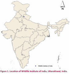
The
following study was conducted at the Chandrabani
Forest Division (30.2830E & 77.9740N), Wildlife
Institute of India campus, Dehradun (Fig. 1).
The study area is ~3.44ha. The
region is characterized with a sub-tropical climate, experiencing cold winters,
warm springs, hot summers and a strong monsoon.
The average annual rainfall received is usually around ~2073.3mm.
The
vegetation is natural and semi-natural represented by a mosaic of natural
scrub, woodland, various successional stages of Shorea
robusta forest including stream bank vegetation
and grassy banks. Thirty-three species
of herpetofauna inhabit the campus; amphibians: 11
species belonging to four families (Bufonidae, Microhylidae, Rhacophoridae, Dicroglossidae), with two species of toads, and reptiles:
22 species belonging to nine families (Colubridae, Typhlophidae, Elapidae, Agamidae, Varanidae, Natricidae, Trionichydae, Geoemydidae, Scincidae) as listed
on the campus database (www.wii.gov.in).
All wildernesses are in close proximity to and in certain parts,
interspersed with human habitation.
The
study was conducted from mid March to the first week of May 2015. Dehradun, having already received its early
showers at the end of February and early March, marked the onset of breeding
activity of Duttaphrynus melanostictus.
Tadpoles started appearing at the natal site by the end of March to
early April.
Methods
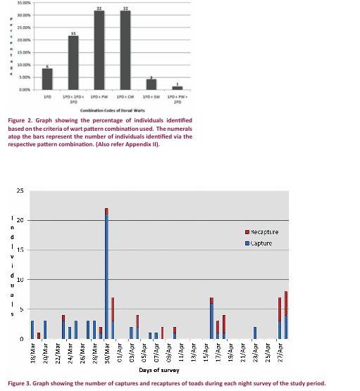
During
the study period (18 March 2015 – 28 April 2015), regular night surveys were
conducted for 42 days. Usually a set of two digital images were generated for each captured
animal, whereby the first image was in portrait mode and the second in
landscape mode. This was done in order
to obtain clear and properly focussed image sets for the dorsal side of each
individual. This also helped in negating
the problems in analysing the wart patterns due to discrepancies in the
position of the animal when being photographed.
If an animal exhibited certain distinct marks (such as deformity or
scar), features or patterns on any part of the body, a third digital image was
generated to showcase the distinctive features. The digital images generated were assigned
unique identification codes, affixed with other data of the animal collected,
and clear black and white photographs were obtained by printing a single
photograph in the complete frame of an A4 sheet. The photographs were then subjected to visual
analysis and manual scrutiny in order to determine the distinctive aspects of
individuals.
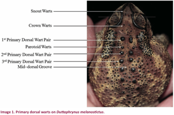
For the
purpose of this study the dorsal warts and associated structures were
classified with respect to the position of their occurrence on toads as follows
(Image 1; Table 1):
Snout
warts: Keratinized small tubercles present in between cranial ridges in front
of upper eyelids. The position and
pattern of distribution with respect to the cranial ridges as well as
arrangements of these warts are used as one character. It is a discontinuous
character as toads may lack tubercles or warts in this region.
Crown
warts: The region of the head from the point where the cranial ridge is notched
along the eye to the anterior end of the parotoid
glands is termed the crown (sensu stricto
Daniel 2002), hence keratinized tubercles present in this region toad are
referred to as crown warts. The position
of these warts with respect to the cranial ridges, parotoid
glands and the first primary dorsal wart pair exhibits great variation, as do
their shapes and patterns of appearance.
This is a discontinuous character, with some toads lacking crown warts.
Mid-dorsal
groove: An associated structure aiding in the classification of dorsal warts
and subsequent identification of individuals.
A distinct dorsal groove is observed along the vertebral axis of the
toad on the dorsal side. It becomes conspicuous along the plane of the anterior
portion parotoid glands, just behind the crown, and
runs all through the entire length of the body up to the vent. The mid-dorsal groove is usually smooth and
usually lacks any tubercles or warts but a few keratinized tubercles might be
found in the groove. The region shows distinct lateral undulations.
Primary
dorsal wart pairs: Two series of large warts along the middle of the dorsal
surface of the toad’s body and exhibiting a certain degree of symmetry on
either side of the distinct mid-dorsal groove.
The primary dorsal warts are considerably enlarged and usually more
keratinized in the adults. The primary
dorsal warts appear to maintain a constant distance from the mid-dorsal
groove. A certain wart of a pair may
often be found associated in close proximity to a small secondary or satellite
wart alongside it. They show great
variability in their position and pattern of distribution. They are commonly found to be oval or
spheroid in shape though some individuals do exhibit a conspicuous shape.
1st
primary dorsal wart pair: It is the first large, distinct and keratinized wart
encountered after the crown warts. It follows just behind the origin of the
mid-dorsal groove, in the hind neck region, and is placed within one-third of
the length of the parotoid glands from the anterior
end. It often exhibits variability in
shape and in the symmetry of its position on either side of the mid-dorsal
groove. A satellite wart is found quite
rarely. It is a continuous character
being observed in all individuals.
2nd
& 3rd primary dorsal wart pair: These are found closely
associated to each other usually towards the distal end with respect to the
plane of the parotoid glands. Variability in shape is less conspicuous and
they usually are spheroid or ovate.
Often found associated with a satellite wart. Varies with respect to position and
pattern. It is a continuous character,
being encountered in all individuals.
Parotoid warts: These warts are encountered along the parotoid
glands on either side of the mid-dorsal groove and in the region between the 1st
and 2nd primary dorsal wart pairs.
They exhibit great variability with respect to their pattern, shape,
position and distribution. It is a
discontinuous character as certain toads don’t bear
these warts.
The above mentioned potential characters may be coded as shown
in Table 1.
The
photographs of the Duttaphrynus melanostictus individuals, obtained during the duration
of the nocturnal surveys at the study site, were subjected to rigorous visual
scrutiny, matching and analysis. We then
attempted to segregate the individual toads based on the combinations of dorsal
warts required to effectively distinguish and identify individuals. This allowed us to arrive at a pattern to be
followed during visual analysis of photographs while sequestering individual
toads to a sub-group.
Table 1. Depicting the dorsal warts and
their coding.
|
Character |
Type |
Code |
|
Snout Warts |
Discontinuous (not encountered in all individuals) |
SW |
|
Crown Warts |
Discontinuous |
CW |
|
1st Primary Dorsal Wart Pair |
Continuous (encountered in all individuals) |
1PD |
|
2nd Primary Dorsal Wart Pair |
Continuous |
2PD |
|
3rd Primary Dorsal Wart Pair |
Continuous |
3PD |
|
Parotoid Warts |
Discontinuous |
PW |
RESULTS
The 1st
primary dorsal wart pair was found to be the initial basis of analyzing the dorsal warts, owing to its apparent consistency
in position and shape. The other dorsal
wart characteristics were now used in combination to the 1st primary
dorsal wart pair to distinguish and identify the individuals. Visual matching and analysis thus led to the
development of a combination of dorsal wart characters based on which the
individuals were subjected to effective individual identification and
subsequent grouping as mentioned in Table 2.
The
photographs of the Duttaphrynus melanostictus individuals, obtained during the duration
of the nocturnal surveys at the study site, were subjected to rigorous visual
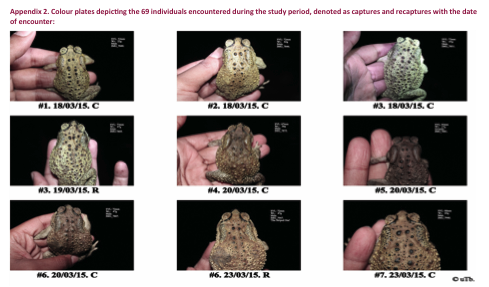
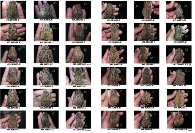
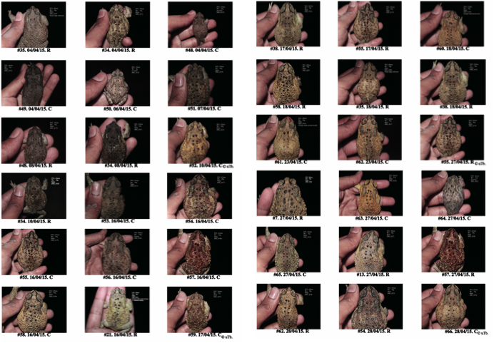
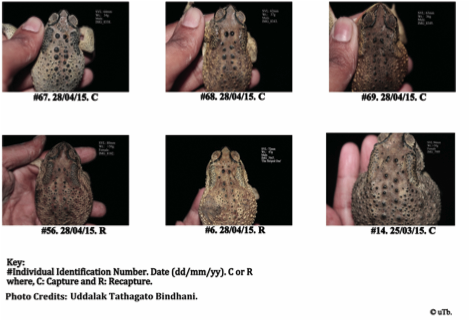
scrutiny, matching and analysis (Appendix 2).
We then attempted to segregate the individual toads based on the
combinations of dorsal warts required to effectively distinguish and identify
individuals. This allowed us to arrive
at a pattern to be followed during visual analysis of photographs while
sequestering individual toads to a sub-group.
Table 2. Dorsal wart pattern
combinations used for individual identification of toads.
|
Combination of dorsal wart characters |
Combination code |
Remarks |
|
1st primary dorsal wart pair
only |
1PD |
Based on variability of shape, size,
pattern, satellite wart and symmetry. |
|
1st primary dorsal wart pair
+ 2nd primary dorsal wart pair + 3rd primary dorsal
wart pair |
1PD + 2PD + 3PD |
Based on variability of satellite warts,
pattern and position. |
|
1st primary dorsal wart pair
+ parotoid warts |
1PD + PW |
Based on variability of pattern,
position and size. |
|
1st primary dorsal wart pair
+ crown warts |
1PD + CW |
Based on variability of pattern,
position and size. |
|
1st primary dorsal wart pair
+ snout warts |
1PD + SW |
Based on variability of pattern and
position. |
|
1st primary dorsal wart pair
+ parotoid warts + 2nd primary dorsal
wart pair |
1PD + PW + 2PD |
Based on variability of shape, size,
pattern, satellite wart and symmetry. |
DISCUSSION
In the
wake of the rising concerns of global decline in amphibian populations (Stuart
et al. 2004; Whittaker et al. 2013) the need of methods and protocols for
sampling natural populations of amphibians has been greatly realized. It is here that mark-recapture techniques of
capturing, marking, releasing and recapturing animals have become an
indispensable tool to monitor and estimate trends in populations. Mark-recapture techniques are advantageous,
being statistically more accurate and robust than uncorrected counts of indices
of relative abundance (Lettink 2012).
Visual
image matching of natural markings is significantly more accurate than invasive
techniques like toe-clipping and computer-assisted image matching, which though
useful for large datasets are constrained by the position and posture of the
animal, glare, shadows, lighting, background colour, equipment and cumbersome
processing protocols to be followed, which can expose animals to prolonged
durations of stress and handling (Caorsi et al. 2012;
Sanchez et al. 2018). Invasive
techniques like toe-clipping, especially for the first
finger, might adversely affect amplexus in males
owing to the loss of the nuptial callosities on phalanges. Thus there is a need for non-invasive
identification techniques, as amphibians are most active during the breeding
season (Sutherland 2006).
Dorsal
warts were found to be a reliable and cheap way to ascertain and monitor
populations in Duttaphrynus melanostictus.
Thus, the technique may also be used in capture-recapture studies of
this species. The study achieved an
accuracy of 100%, whereby the digital image sets of the toads successfully
distinguished and identified all 69 individuals (Appendix 1).
The
results indicate that it should be possible to efficiently process photographs
of unidentified captures in a full-scale monitoring programme by using the
combination code key to identify and determine the identity of any given
capture.
Analysis
of photographs of 69 toads identified six combinations which
resulted in optimal allocation of individuals into captures and
recaptures. The decision as to which of
these combinations to use in future studies shall depend on the clarity of the
photographs of the dorsal side of the toad taken in the field. It is recommended that the 1st
primary dorsal wart pair should be considered initially.
The 2nd
primary dorsal wart pair was always found to be in symmetry, pattern and
variation with the 3rd primary dorsal wart pair or the parotoid warts if present.
Thus, establishment of individual identity was never made based on the 2nd
and 3rd primary dorsal wart patterns alone, and thus were considered
a separate combined character combination with the 1st primary
dorsal wart pair.
It
might also be mentioned that there were eight toads (~ 11.59%) that also
exhibited certain distinct marks, patterns, wounds or infections. Preliminarily these could be used as a cue
for individual identification, especially when in the field, complementing the
dorsal wart patterns. But, it was seen
that there was no constancy (wounds and infections heal, body marks might be
lost during moulting and sloughing of skin etc.) of these characters, and thus
they are unsuitable for application to individual identification in the long
run.
This synchronized scientific method is simple
to follow and easy to implement, and thus can even be utilized by laymen in the
field of biology to monitor toads in their backyards. The study also holds great value, both
scientific and economic, in keeping tabs of toad populations threatened from
road related mortality. It thus shows
great potential to be successfully utilized and implemented in citizen science
programmes aimed at studying amphibians.
REFERENCES
Begon, M. (1979). Investigating Animal Abundance:
Capture-recapture for Biologists. Edward Arnold
(Publishers) Ltd., 97pp.
Bradfield, K.S. (2004). Photographic
Identification of Individual Archey’s Frogs, Leiopelma archeyi, from Natural
Markings. Department of Conservation, Wellington,
New Zealand, 36pp.
Caorsi, V.Z., R.R. Santos & T. Grant (2012). Clip or snap? An
evaluation of toe-clipping and photo-identification methods for identifying
individual Southern Red-Bellied Toads, Melanophryniscus
cambaraensis. South
American Journal of Herpetology 7(2): 79–84.
Caughley, G. &
A.R.E. Sinclair (1994). Wildlife Ecology and Management. Blackwell
Science, Oxford, 334pp.
Daniel, J.C. (2002). The Book of
Indian Reptiles and Amphibians. Bombay Natural
History Society and Oxford University Press, Bombay, 252pp.
Daniels, R.J. (1994). Dorsal warts identify individual common
Indian toads. Cobra 16: 22–24.
Daniels, R.R. (2005). Amphibians of
peninsular India. Universities Press, India,
337pp.
Donnelly, M.A., C. Guyer,
J.E. Juterbock
& R.A. Alford (1994). Techniques for marking amphibians; pp 277– 284. In: Heyer,
W.R., M.A. Donnelly, R.W. McDiarmid, L-A.C.
Hayek & M.S. Foster (eds.).
Measuring and Monitoring Biological Diversity: Standard Methods for Amphibians.
Smithsonian University Press, Washington DC, 384pp.
Heyer, W.R., M.A. Donnelly, R.W. McDiarmid, L-A.C. Hayek & M.S. Foster (eds.) (1994). Measuring and Monitoring Biological
Diversity: Standard Methods for Amphibians. Smithsonian University Press, Washington
DC.
Lettink, M. (2012). Herpetofauna:
population estimates (using capture - mark - recapture data) Version 1.0.
Inventory and monitoring toolbox: herpetofauna. Department
of Conservation, Govt. Of New Zealand, 27pp.
Sanchez, E., S. Gippner,
M. Vences, K. Preißler,
I.J. Hermanski, B.A. Caspers,
E.T. Krause, S. Steinfartz & F.W. Kastrup (2018). Automatic quantification of color proportions in dorsal black and yellow colored amphibians, tested on the Fire Salamander (Salamandra salamandra).
Herpetology Notes 11: 73–76.
Stuart, S.N., J.S. Chanson, N.A. Cox, B.E.
Young, A.S. Rodrigues, D.L. Fischman & R.W.
Waller (2004). Status and trends of amphibian declines and extinctions worldwide. Science
306(5702): 1783–1786; https://doi.org/10.1126/science.1103538
Sutherland, W.J. (ed.). (2006). Ecological Census Techniques: A
Handbook. Cambridge University Press, United Kingdom,
448pp.
Whittaker, K., M.S. Koo, D.B. Wake &
V.T. Vredenburg (2013). Global declines of amphibians, pp.
691–699. In: Levin, Simon A. (eds.) Encyclopedia
of Biodiversity: Vol III. Academic Press, 5504pp;
https://doi.org/10.1016/B978-0-12-384719-5.00266-5
Appendix 1. Data sheet table, as
prepared, for the creation of dorsal wart pattern combination codes and thus,
distinctly identifying individuals of D. melanostictus
from the Wildlife Institute of India campus, where ‘1’ represents the
character being used and ‘0’ represents that the character wasn’t utilized for
identifying the individual:
|
Individual Id. |
1 – PDW |
2 – PDW |
3 – PDW |
SW |
CW |
PW |
|
#1. |
1 |
0 |
0 |
0 |
0 |
0 |
|
#2. |
1 |
0 |
0 |
0 |
0 |
1 |
|
#3. |
1 |
0 |
0 |
0 |
1 |
0 |
|
#4. |
1 |
0 |
0 |
0 |
0 |
1 |
|
#5. |
1 |
1 |
1 |
0 |
0 |
0 |
|
#6. |
1 |
0 |
0 |
0 |
1 |
0 |
|
#7. |
1 |
0 |
0 |
0 |
1 |
0 |
|
#8. |
1 |
0 |
0 |
0 |
0 |
1 |
|
#9. |
1 |
0 |
0 |
0 |
0 |
1 |
|
#10. |
1 |
0 |
0 |
0 |
0 |
0 |
|
#11. |
1 |
0 |
0 |
0 |
0 |
1 |
|
#12. |
1 |
0 |
0 |
0 |
0 |
1 |
|
#13. |
1 |
0 |
0 |
0 |
1 |
0 |
|
#14. |
1 |
0 |
0 |
0 |
0 |
1 |
|
#15. |
1 |
0 |
0 |
0 |
1 |
0 |
|
#16. |
1 |
0 |
0 |
0 |
0 |
1 |
|
#17. |
1 |
1 |
1 |
0 |
0 |
0 |
|
#18. |
1 |
1 |
1 |
0 |
0 |
0 |
|
#19. |
1 |
0 |
0 |
0 |
0 |
1 |
|
#20. |
1 |
1 |
1 |
0 |
0 |
0 |
|
#21. |
1 |
0 |
0 |
0 |
1 |
0 |
|
#22. |
1 |
1 |
1 |
0 |
0 |
0 |
|
#23. |
1 |
0 |
0 |
0 |
1 |
0 |
|
#24. |
1 |
0 |
0 |
0 |
1 |
0 |
|
#25. |
1 |
0 |
0 |
0 |
0 |
0 |
|
#26. |
1 |
0 |
0 |
0 |
1 |
0 |
|
#27. |
1 |
0 |
0 |
0 |
1 |
0 |
|
#28. |
1 |
0 |
0 |
0 |
1 |
0 |
|
#29. |
1 |
0 |
0 |
0 |
0 |
1 |
|
#30. |
1 |
0 |
0 |
0 |
0 |
1 |
|
#31. |
1 |
1 |
1 |
0 |
0 |
0 |
|
#32. |
1 |
0 |
0 |
1 |
0 |
0 |
|
#33. |
1 |
1 |
1 |
0 |
0 |
0 |
|
#34. |
1 |
1 |
0 |
0 |
0 |
1 |
|
#35. |
1 |
1 |
1 |
0 |
0 |
0 |
|
#36. |
1 |
1 |
1 |
0 |
0 |
0 |
|
#37. |
1 |
0 |
0 |
0 |
0 |
1 |
|
#38. |
1 |
0 |
0 |
0 |
1 |
0 |
|
#39. |
1 |
1 |
1 |
0 |
0 |
0 |
|
#40. |
1 |
1 |
1 |
0 |
0 |
0 |
|
#41. |
1 |
0 |
0 |
0 |
1 |
0 |
|
#42. |
1 |
0 |
0 |
0 |
1 |
0 |
|
#43. |
1 |
0 |
0 |
0 |
0 |
1 |
|
#44. |
1 |
1 |
1 |
0 |
0 |
0 |
|
#45. |
1 |
0 |
0 |
0 |
0 |
1 |
|
#46. |
1 |
0 |
0 |
0 |
1 |
0 |
|
#47. |
1 |
0 |
0 |
0 |
0 |
1 |
|
#48. |
1 |
1 |
1 |
0 |
0 |
0 |
|
#49. |
1 |
0 |
0 |
0 |
1 |
0 |
|
#50. |
1 |
0 |
0 |
0 |
1 |
0 |
|
#51. |
1 |
0 |
0 |
0 |
0 |
1 |
|
#52. |
1 |
0 |
0 |
0 |
1 |
0 |
|
#53. |
1 |
0 |
0 |
1 |
0 |
0 |
|
#54. |
1 |
0 |
0 |
0 |
0 |
1 |
|
#55. |
1 |
0 |
0 |
0 |
0 |
0 |
|
#56. |
1 |
0 |
0 |
0 |
0 |
1 |
|
#57. |
1 |
1 |
1 |
0 |
0 |
0 |
|
#58. |
1 |
0 |
0 |
0 |
1 |
0 |
|
#59. |
1 |
0 |
0 |
0 |
0 |
1 |
|
#60. |
1 |
0 |
0 |
1 |
0 |
0 |
|
#61. |
1 |
0 |
0 |
0 |
0 |
1 |
|
#62. |
1 |
0 |
0 |
0 |
0 |
1 |
|
#63. |
1 |
0 |
0 |
0 |
1 |
0 |
|
#64. |
1 |
0 |
0 |
0 |
0 |
0 |
|
#65. |
1 |
0 |
0 |
0 |
1 |
0 |
|
#66. |
1 |
1 |
1 |
0 |
0 |
0 |
|
#67. |
1 |
0 |
0 |
0 |
1 |
0 |
|
#68. |
1 |
0 |
0 |
0 |
0 |
1 |
|
#69. |
1 |
0 |
0 |
0 |
0 |
0 |